

How the Circulatory System Works: Heart to Cells
On this page· 17
You're sitting still, reading this, and your blood isn't. Right now it's leaving your heart, fanning out to your eyes, fingers and feet, swapping oxygen for waste in vessels too fine to see, then heading back to start again. You don't have to think about any of it, which is why most of us carry a fuzzy picture of how the circulatory system works, something like a pump pushing liquid around pipes.
That picture is a decent start, but it hides the interesting parts. Your heart is really two pumps in one, your veins hold most of your blood, and tiny vessels near your cells decide, moment by moment, which tissues get more. Follow one red blood cell on its route and the whole system starts to make sense.
The Two Circuits at a Glance
Blood follows two connected routes. The right side of the heart sends it to the lungs; the left side sends it around the body. Like two delivery loops sharing one depot, they keep blood moving between gas exchange in the lungs and the tissues that use oxygen.
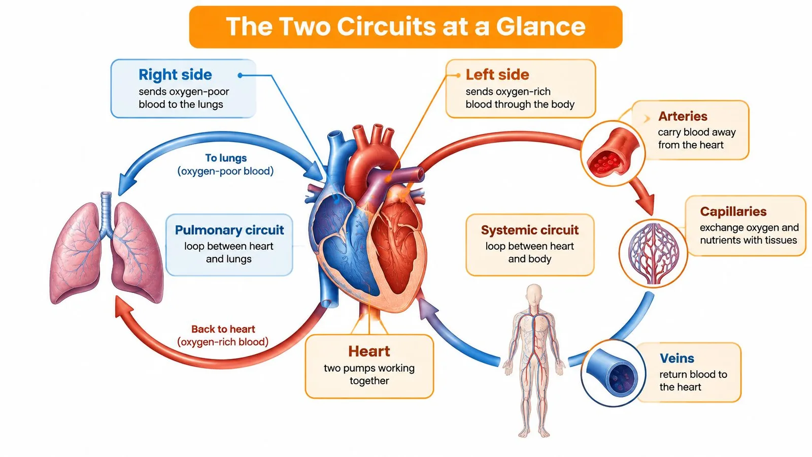
On the body route, called the systemic circuit, blood leaves the left side of the heart through arteries. These branch into smaller vessels and then into capillaries, where oxygen and nutrients pass into tissues while carbon dioxide and other wastes enter the blood, as described in the OpenStax chapter on the cardiovascular system. Veins collect the blood and return it to the heart. Valves in larger veins help keep it moving toward the heart despite low pressure and gravity.
One Red Blood Cell, One Full Lap
A red blood cell leaves the left ventricle through the aorta, travels through branching arteries, and reaches a capillary in a muscle such as the calf. There, it releases oxygen and picks up carbon dioxide. Small veins merge into larger ones, carrying the cell to the right atrium.
From there, the route continues through the pulmonary circuit. The right ventricle sends blood to the lungs, where it releases carbon dioxide and takes up oxygen. The blood then returns to the left atrium and passes into the left ventricle, ready for another trip around the body.
The map in one line: The heart drives blood forward, arteries distribute it, capillaries exchange materials, and veins return it.
| Circuit | Route of blood | Main job |
|---|---|---|
| Systemic circuit | Left ventricle, aorta, arteries, capillaries, veins, right atrium | Delivers oxygen and nutrients to body tissues and collects carbon dioxide and waste |
| Pulmonary circuit | Right ventricle, pulmonary arteries, lungs, pulmonary veins, left atrium | Picks up oxygen and releases carbon dioxide in the lungs |
Systemic veins hold most of the body's blood at any moment. This reserve lets circulation shift blood toward tissues that suddenly need more, rather than requiring the heart to produce extra blood. For more clinical detail on the two loops, the Ace Med Boards cardiovascular physiology guide explains their pressures and control.
Question 1 of 5
Why is the muscular wall on the right side of the human heart so much thinner than the wall on the left?
How the Heart Pumps Blood Around the Body
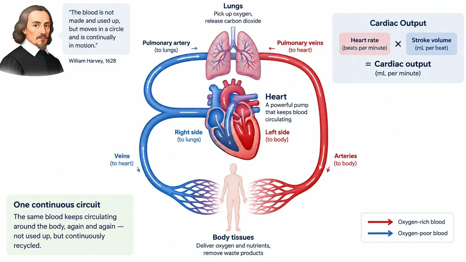
That blood moves in a closed circuit at all was once a radical claim. English physician William Harvey made it in his 1628 book De Motu Cordis, arguing that blood circulates continuously rather than being consumed and remade, as the Britannica biography of William Harvey describes. Harvey estimated how much blood the heart must move and showed the quantity was far too large for tissues to use up.
Four Chambers, Four One-Way Doors
The right atrium and right ventricle form the low-pressure pump for the lungs. The left atrium and left ventricle form the higher-pressure pump for the body, which is why the left ventricle wall is thicker. It has a much longer route to supply.
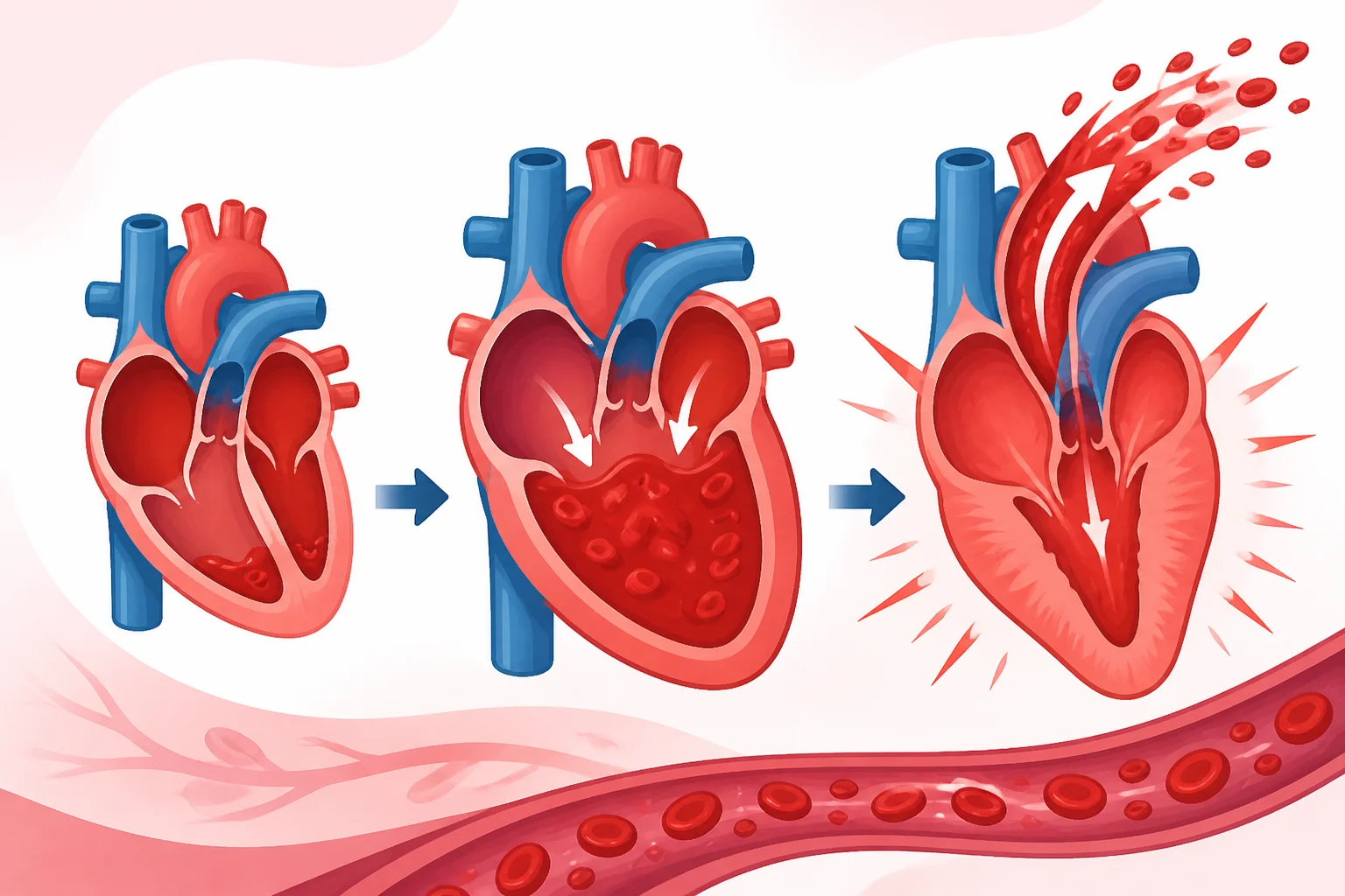
Blood meets four valves in order. The tricuspid valve lets it into the right ventricle, the pulmonary valve lets it out to the lungs, the mitral valve lets it into the left ventricle, and the aortic valve lets it out to the body. Each opens forward and snaps shut, so a squeeze moves blood onward instead of backward.
The Throughput Formula
How much blood does this pump move? The StatPearls chapter on cardiac output in the NCBI Bookshelf defines cardiac output as heart rate multiplied by stroke volume, written as CO = HR × SV. At rest, a healthy adult typically pumps about 4 to 6 L/min, and during severe exercise output can rise to roughly three- to fourfold.
| Situation | Heart rate | Stroke volume | Cardiac output |
|---|---|---|---|
| Resting healthy adult | about 70 beats per minute | about 70 mL per beat | about 4 to 6 L/min |
| Vigorous exercise, trained adult | about 180 beats per minute | about 140 mL per beat | roughly 25 L/min |
| Very fast heart rate with short filling time | higher | can fall | may plateau or fall |
A Pump That Adjusts Itself
The heart also self-corrects, beat by beat. When more blood returns and fills a ventricle, the stretched muscle contracts more forcefully and ejects a larger stroke. That's the Frank-Starling mechanism, a passive mechanical response, not a fresh nerve signal, and it stops blood from backing up in the veins when inflow rises.
Here's the trade-off that catches people out. A faster heart rate doesn't always raise output, because very fast beats shorten filling time, stroke volume falls, and the product of the two can stall. Output depends on filling, contractility, and the resistance the ventricle pushes against, all matched to what your tissues are asking for.
Question 2 of 5
Why does a heart that fills with more blood automatically squeeze harder on the next beat?
Arteries, Capillaries and Veins Explained
Roads make a fair analogy here, as long as you remember these roads can widen and narrow on their own. Arteries are the high-pressure highways leaving the heart. Arterioles are the controllable junctions that decide how much traffic each neighborhood gets. Capillaries are the narrow delivery streets where goods are handed over, and veins are the return roads home.
Walls Built for the Job
Arteries handle blood arriving in pulses with each heartbeat, so their walls are thick, elastic and muscular. They stretch slightly with each surge and recoil between beats, which smooths flow downstream. Capillaries sit at the opposite extreme, with walls only one cell thick, because oxygen, glucose and carbon dioxide have to cross quickly and over a very short distance.
| Vessel type | Direction of flow | Pressure level | Key structure |
|---|---|---|---|
| Arteries | Away from the heart | High and pulsing | Thick elastic and muscular walls |
| Arterioles | Into capillary beds | Falling; main adjustable resistance | Muscular walls that change diameter |
| Capillaries | Exchange zone between blood and tissue | Low and steady | Walls only one cell thick |
| Venules and veins | Back toward the heart | Low | Valves in larger veins prevent backflow |
Arterioles Do the Directing
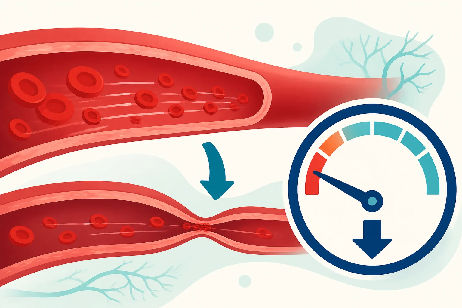
The heart sets overall flow capacity, but arterioles allocate it. A review in PubMed Central on systemic circulation explains that vascular resistance is approximately proportional to 1/r⁴ under idealized laminar-flow conditions. In plain terms, halving a vessel's radius doesn't halve flow, it cuts it far more sharply, so small changes in arteriole diameter produce large changes in delivery.
Tissues exploit this. When a muscle works hard, local signals such as reduced oxygen and increased carbon dioxide relax its arterioles, resistance drops, and more blood arrives without the heart having to work proportionally harder. Quieter tissues get less. It's demand-led distribution, handled locally.
The heart decides how much blood is available. Arterioles decide where it goes.
The Low-Pressure Trip Home
By the time blood reaches the veins, most of the pressure from the heartbeat has been spent. Veins are compliant and stretchy, which makes them a useful reservoir, but returning blood uphill from your legs needs help. Valves prevent backflow, and contracting leg muscles squeeze the veins, nudging blood toward the heart each time you walk. That's why standing motionless for hours leaves legs heavy, while walking relieves them.
Question 3 of 5
Why does a tiny narrowing of an arteriole cause a much bigger drop in blood flow than you'd expect?
Pressure Is a Readout, Not the Push
A cuff reading captures pressure in a large artery, but it does not show how well blood reaches every tissue. Blood flow depends on the heart's energy and on factors such as blood volume, vessel elasticity, vessel shape, and resistance, as described in this physiology review of what makes blood move. Pressure is one readout of how those parts are working together.
Veins show why pressure alone cannot explain circulation. Their pressure is low, yet blood returns to the heart because stretchy vein walls hold blood, while muscle movement, breathing, and heart function help move it along. A low reading in one part of the route does not mean blood has stopped moving.
Three Linked Numbers, Not One
Physiologists summarize the relationship as CO ≈ MAP/TPR: cardiac output approximately equals mean arterial pressure divided by total peripheral resistance. The equation shows how flow and resistance shape pressure. When arterioles constrict, resistance rises and pressure can increase even as flow to a particular tissue falls. When arterioles widen, local blood supply can improve without a rise in overall pressure.
Higher blood pressure does not automatically mean better circulation. High blood pressure and cardiovascular disease are among the leading causes of death worldwide. Tissues need blood flow matched to their needs, not the highest possible pressure. Some familiar ideas about the body need correcting, as these common science misconceptions illustrate.
When Normal Numbers Hide a Local Problem
A blood pressure cuff measures the macrocirculation in larger vessels. The microcirculation describes blood flow through tiny vessels in tissue, where local supply can be impaired even when blood pressure or cardiac output appears acceptable. Fingertip skin blood-flow measurements have been found to correlate with capillary refill time and lactate, but not necessarily with cardiac output.
Cold fingers offer an everyday example. Skin arterioles constrict to conserve heat, so fingers can turn pale and numb while the body's core stays warm. A runner shows the opposite distribution: arterioles widen in working leg muscles and narrow in the gut.
- Pressure: A cuff's reading in a large artery. It is useful, but does not show the whole route.
- Flow: The amount of blood moving through a vessel or tissue per minute.
- Delivery: Whether blood supplies oxygen to cells in a particular capillary bed.
In shock, sepsis, and diabetes-related vessel damage, tissue flow can fall while large-vessel readings remain acceptable. Clinicians therefore also assess signs such as capillary refill, skin mottling, and temperature, and may use Doppler or optical measurements. A normal pulse or blood pressure cannot confirm that every tissue is adequately supplied. Nor does a cold hand at home, by itself, diagnose a disease.
Read circulation as a route, not a single number: pressure reflects conditions in larger vessels, flow varies with tissue demand, and delivery depends on blood reaching the cells. The heart and vessels must work together for that delivery to happen.
Question 4 of 5
Why can blood pressure rise even while less blood reaches a particular tissue?
How Oxygen and Nutrients Reach Every Cell
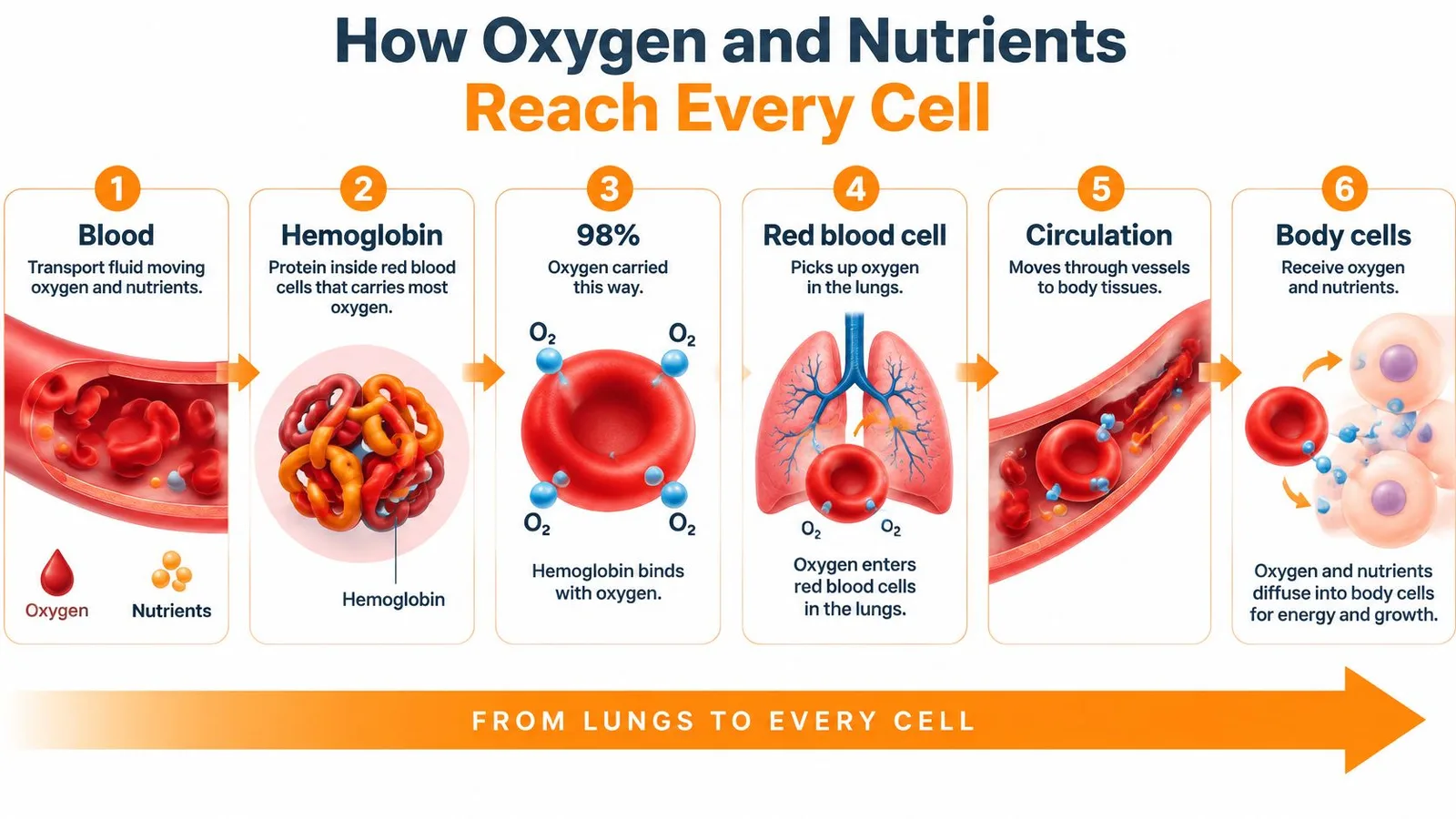
Blood itself is the transport fluid, and red blood cells are its cargo carriers. Inside each one sits hemoglobin, a protein that grabs oxygen where it's plentiful and releases it where it's scarce. Most of the oxygen in your blood rides this way, bound to hemoglobin rather than dissolved freely, which vastly increases how much each liter can carry.
Six Stops for One Red Blood Cell
- Loading in the lungs: Oxygen diffuses from air sacs into capillary blood and binds to hemoglobin, while carbon dioxide moves out to be exhaled. Loading here works because oxygen is abundant in freshly inhaled air.
- Launch from the left ventricle: Oxygen-rich blood is pushed into the aorta, giving it the energy to reach the body's farthest tissues.
- Routing at the arterioles: Junction vessels steer more blood toward active muscle and less toward resting areas, so supply tracks demand instead of spreading evenly.
- Exchange in the capillaries: Oxygen leaves hemoglobin, crosses the one-cell-thick wall, and enters tissue, while glucose and other nutrients move out and carbon dioxide and wastes move in.
- The return load: Venous blood carries carbon dioxide back partly dissolved, partly as bicarbonate, and partly bound to hemoglobin. Splitting the load keeps blood chemistry stable on the trip.
- Unloading in the lungs: The right ventricle sends this blood to the lungs, carbon dioxide is exhaled, and the cell reloads with oxygen for the next lap.
Where the Picture Goes Wrong
Many readers imagine blood dumping oxygen into an organ in bulk, like emptying a bucket. Exchange doesn't work that way. It happens across thin capillary walls, one small zone at a time, which is why capillary networks are so dense that nearly every cell sits close to one.
Delivery rule: If capillaries can't be reached or their walls and flow are damaged, tissue suffers even when large vessels look open.
That local step is exactly where chronic conditions bite. In diabetes, damaged small vessels can starve skin and underlying tissue of the oxygen needed for repair, and clinicians who describe vascular care for non-healing diabetic wounds focus on restoring that last stretch of delivery. For more surprising details about transport and other body systems, browse these interesting facts about the human body and test which ones match the route you've just traced.
Question 5 of 5
What makes hemoglobin let go of its oxygen right where a hard-working muscle needs it?
When to Seek Medical Care
Chest discomfort, breathlessness, fainting, or sudden changes in a limb can signal that blood is not reaching tissues adequately. The symptoms cannot identify the cause on their own, but they can tell you how quickly to seek help.
Call emergency services now for chest pain or pressure, shortness of breath at rest, or fainting. Call as well if skin becomes blue, cold, or very pale, especially with breathlessness or chest discomfort. Don't wait for a routine appointment or drive yourself. A sudden change in skin color or temperature, or sudden swelling in one leg without those emergency symptoms, needs same-day medical evaluation.
A clinician can assess circulation by checking blood pressure and pulse alongside tissue perfusion, capillary refill, skin condition, temperature, and laboratory markers. A home reading cannot assess all of these, and a normal pulse or blood pressure does not rule out a local delivery problem. Care after cardiac arrest can involve optimizing ventilation after return of circulation.
Heat can also strain circulation by drawing blood toward the skin. Learn the signs that distinguish heat stroke from heat exhaustion. If you have chest discomfort, breathlessness at rest, fainting, or blue, cold, or very pale skin, call emergency services now. For a sudden change in skin color or temperature, or sudden swelling in one leg without those emergency symptoms, seek same-day medical evaluation.
Million Whys turns everyday curiosity into short questions with explanations for every answer choice, including incorrect ones. Try a daily question, follow a topic further, and build a personal map of what you've learned.
Keep Exploring
Related Posts
How Do Batteries Work: A Simple Guide to the Science
Learn how do batteries work, from the chemistry inside each cell to voltage, capacity, and the real factors that shape everyday performance.
Facts About Nature: Hidden Systems That Keep Life Moving
Facts about nature get better when you see the hidden systems: water cycling, carbon moving, fungi trading, pollinators connecting, and decay renewing life.
Fun Facts About Human Body: Everyday Biology
Fun facts about human body biology: why blinking, heart rate, gut rumbles, body clocks, and sneezing work as tiny automatic survival systems inside you.